Imaging Contrast Media
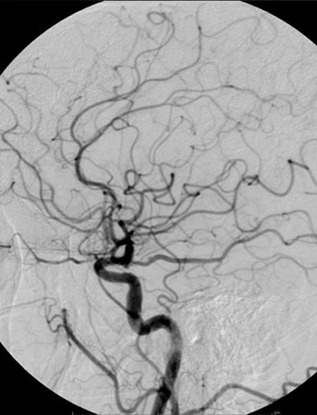
Contrast materials, also called contrast agents or contrast media, are used to improve pictures of the inside of the body produced by x-rays, computed tomography (CT), magnetic resonance (MR) imaging, and ultrasound. Often, contrast materials allow the radiologist to distinguish normal from abnormal conditions. Contrast materials are not dyes that permanently discolor internal organs. They are substances that temporarily change the way x-rays or other imaging tools interact with the body. When introduced into the body prior to an imaging exam, contrast materials make certain structures or tissues in the body appear different on the images than they would if no contrast material had been administered. Contrast materials help distinguish or “contrast” selected areas of the body from surrounding tissue. By improving the visibility of specific organs, blood vessels or tissues, contrast materials help physicians diagnose medical conditions.
(or contrast medium) is a substance used to increase the contrast of structures or fluids within the body in medical imaging.[1] Contrast agents absorb or alter external electromagnetism or ultrasound, which is different from radiopharmaceuticals, which emit radiation themselves. Contrast agents, enhance the radiodensity in a target tissue or structure.
Contrast agents are commonly used to improve the visibility of blood vessels and the gastrointestinal tract. Several types of contrast agent are in use in medical imaging and they can roughly be classified based on the imaging modalities where they are used. Most common contrast agents work based on X-ray attenuation and magnetic resonance signal enhancement.
